Download Detection of Urinary Tract Proteins: Tamm-Horsfall Mucoprotein and Others in Normal Urine and more Study notes Pathology in PDF only on Docsity!
J. cliit. Path. (1959),^ 12,^ 510.
..THE PROTEINS OF^ NORMAL^ URINE
II. FROM THE^ URINARY^ TRACT^ *
BY
GREGOR H. GRANT
WITH THE TECHNICAL ASSISTANCE^ OF^ PHILIP^ H.^ EVERALL
From the Department of^ Pathology,^ the^ Royal Salop^ Infirmary,^ Shrewsbury
(RECEIVED FOR^ PUBLICATION^ JUNE^ 26, 1959)
Normal urine contains traces of many different proteins. These^ proteins^ include:
(1) Plasma^ proteins^ entering^ the urine from^ the
blood through the^ glomeruli.
(2) Urinary tract proteins from the^ glands^ or shed cells of the urinary tract from^ the kidney
tubules downwards.^ One^ protein,^ the^ mucoprotein
ot Tamm and Horsfall (1952), has been described as from^ this^ source. (3) In the male, seminal proteins from^ the^ genital tract and in particular from the^ prostate^ and bulbo-urethral glands, which open directly^ into the urethra. Several proteins have been described^ in seminal plasma (Ross, Moore, and Miller,^ 1942).
Immunochemical methods of analysis have
made it possible not only to demonstrate such proteins in^ normal^ urine,^ but^ even^ to^ investigate their origins. The detection in normal urine^ of most of the plasma proteins (exceptions include fibrinogen and (^) fl-lipoprotein) was^ described
previously (Grant, 1957): evidence for^ the
presence of proteins from other sources is^ now presented.
Materials and Methods Urines Examined.-Individual^ and^ pooled^ samples from four normal men, four normal women, and five children, three boys and two girls, aged 4 to^ 11, were investigated. They were all^ members^ of^ the laboratory staff or^ of^ their^ families.^ Simple^ pre- cautions were^ taken^ to^ avoid^ contamination,^ and^ the samples were preserved with sodium azide (1 g. per litre). They were concentrated by ultrafiltration under negative pressure through^ Visking^ dialysis tubing as^ previously described,^ but,^ instead^ of^ tubing of 3 in. flat width, longer lengths of the narrower in. flat width tubingt were used because of its *Based on a paper read at the combined meeting of the Association of Clinical Pathologists and of the Association of Clinical Biochemists in London on October 4, 1958.^ Part I^ was read at a meeting of the Association of^ Clinical^ Pathologists^ at Torquay on October 12, 1957 (J. cln. Path.,^ 1957,^ 10,^ 360). tObtainable from Hudes Merchandising Corporation,^ 52, Gloucester Place, London, W.I.
ability to withstand^ the^ pressure^ without^ support (Everall and Wright, 1958).^ After the^ urine^ had been
concentrated about^50 times,^ it^ was^ centrifuged^ and
the supernatant was^ further^ concentrated^ in^ a^ fresh length of dialysis tubing^ to^ give^ a^ final^ concentration
of about 1,000 times.^ If^ it is^ concentrated^ in^ a^ single
stage, the final^ product^ is^ often^ too^ viscous^ to^ allow any precipitate to^ settle.
Tamm-Horsfall Mucoprotein.-This^ was^ prepared
from normal^ urine^ (preserved^ with^ sodium azide,^1 g. per litre) by adding^ an^ equal^ volume^ of^ 1.16M
NaCl. The^ precipitate^ was^ washed^ twice^ with 0.58M
saline, and^ then^ dialysed^ against^ distilled^ water containing 1 g.^ per^ litre sodium^ azide, until^ tests^ for chloride became negative. Before analysis it^ was dialysed against^ the^ appropriate^ buffer. Such preparations when freshly made^ will^ not diffuse through agar even in an electric field.^ On standing, however, they become in time^ freely diffusible and suitable for use in immunochemical agar gel tests.
Semen,-Samples were^ available^ from^ patients^ who
were referred^ to^ the^ laboratory from^ the^ infertility clinic and^ subsequently^ found^ to^ be^ normal.
Antisera.-These were prepared in^ rabbits against
(1) concentrated normal male urine^ colloids; (2)^ con- centrated normal female urine^ colloids;^ (3)^ Tamm- Horsfall mucoprotein freshly^ prepared^ as^ above; and (4) normal human semen. At least two rabbits were used for each; the courses of injection were as previously described; and^ the
antisera were preserved with sodium^ azide^ (1^ in^ 1,000).
Before use antibodies^ against^ serum^ proteins^ were removed by absorption. This absorption proved to be unexpectedly difficult, for the antisera were found to contain^ antibodies against proteins which^ are^ present^ in^ exceedingly^ low concentrations in normal human serum. Human
serum, concentrated four times by the same technique
as that used for concentrating urine^ colloids,^ was therefore used^ for^ their^ absorption,^1 volume^ of
concentrate to 10 volumes of antiserum. The
absorbed antisera then gave no precipitin reaction with an equal volume of^ human serum^ concentrate,
THE PROTEINS OF NORMAL URINE: PART 11
an equal volume of fresh serum, or 1/20 volume of fresh (^) serum. In (^) addition to the above antisera, rabbit antisera against normal human serum (^) proteins were required for the analyses of normal human serum run in parallel with the urine analyses to indicate the relative mobilities of the serum and urinary tract proteins.
Immunochemical Analyses.-For the simple agar gel precipitin tests (Ouchterlony, 1949) Petri dishes containing pH 7.4 phosphate agar were used (Gell, 1955). Then 27.5 g. potassium dihydrogen phosphate, 17.0 g. sodium chloride, 8.0 g. potassium hydroxide, and (^) 15 g. agar were (^) dissolved in 2 litres of distilled water and filtered while hot (^) through a bed (^) of "hyflo super cell " under reduced pressure. Sodium azide was added to give a final concentration of (^1) g. per litre and the agar sterilized at 5 lb. pressure for 15 minutes. The immuno-electrophoretic analyses were carried out by the method of Grabar and Williams (1955) modified as previously described (Grant, 1957).
Results
Analyses with Antisera against Normal Urine
Colloids Absorbed with Normal Serum.-These
antisera should demonstrate every urine protein
except those in blood serum.
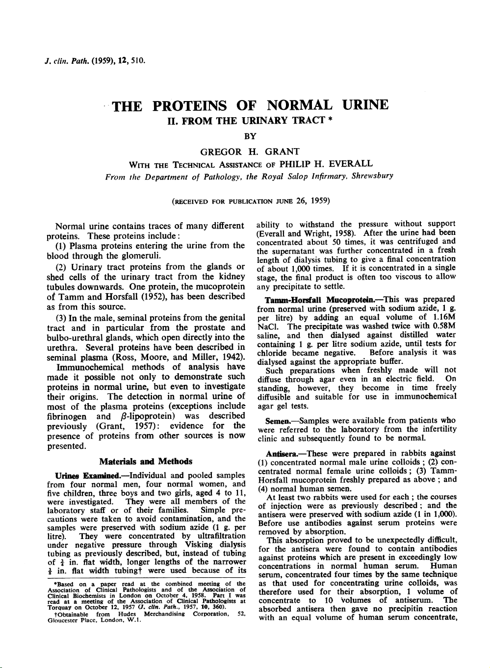
Simple Difflusion-precipitin Analyses.-Fig. 1
illustrates such an analysis; two samples of male
urine concentrate, two samples of female urine
concentrate, and some salt-precipitated Tamm-
Horsfall mucoprotein have been allowed to react
FIG. (^) 1.-Comparison between non-serum proteins of male urine, female urine, and Tamm-Horsfall mucoprotein in (^) peripheral cups. In the centre cup is shown antiserum against normal male urine colloids (absorbed with normal human serum). l(Direct print of Ouchterlony plate stained with azocarmine.)
FIG. (^) 2.-Comparison between male (M.U.), female urine colloids (F.U.), and semen (Se), using the same antiserum. (Similar direct print.) with (^) arl antiserum made against male urine colloids, prepared and absorbed as described above. The urine (^) concentrates form multiple precipitin lines, two of which (^) are particularly well marked. One of the latter gives the " (^) reaction of identity" with the mucoprotein preparation. Similar results were obtained with the antisera made against female urine concentrates. In Fig. 2 urine concentrates have been com- pared with normal semen using a similar antiserum. As expected the antiserum contains antibodies to several seminal proteins and these proteins are only minor (^) components of the urine protein; their precipitin lines cut (^) those of the main urinary tract proteins, including that due to the Tamm-Horsfall mucoprotein, a protein evidently absent from semen.
Similar analyses, using also antisera made
against female urine concentrates, led to a further and (^) unexpected finding, namely, several trace components (^) common to semen and to female urine as well (^) as to (^) male urine. On the basis of
these preliminary experiments three groups of
components can be (^) distinguished:
(1) Components common to male and female
urine but (^) not present in semen: these presumably arise from the (^) upper urinary tract; they include the mucoprotein of Tamm and Horsfall as well as
the other principal components.
(2) Components common only to male urine and semen: (^) they are apparently only present in traces
and presumably arise from the genital tract.
51I
THE PROTEINS OF NORMAL URINE: PART 11
the urine presumably arise from the urethra or
male genital tract: they form the second and third
groups of components classified in the preliminary
experiments above.
Simple Diffusion-precipitin Analyses.-In^ Fig.^6
the use of this type of antiserum to compare the antigenic components of male urine, female urine, and semen is illustrated. It confirms that there are about four components common to semen and to
female urine as well as to male urine. The absence
of the Tamm-Horsfall mucoprotein from semen was also confirmed.
FIG. 5.-Identificationofcomponent antigenicallysimilar to Tamm-Horsfall mucoprotein. U.=male urine concentrate. Left trough= antiserum against male urine colloids absorbed with serum and semen. Right trough=antiserum to the salt-precipitated protein of Tamm and Horsfall.
analyses as that illustrated on the left of Fig. 5.
Comparison with^ serum^ proteins^ analysed^ on^ the
same plate allows a rough estimate of the
mobilities of these urine proteins using the data
of Williams and Grabar (1955).
Of the six precipitin lines, that labelled^ T.H.^ in
Fig. 4 was predominant in all^ analyses. The^ line
AB and occasionally the line G were nearly as
dense with certain antisera. The (^) remaining lines
were always faint.
The line T.H. corresponds to the salt-
precipitated mucoprotein of Tamm and Horsfall
(Fig. 5). It has two humps with maxima^ as shown and sometimes it extends faintly to^ a position opposite the cup, probably owing to
adsorption to material^ remaining unmoved.
Analyses with Antisera against Normal^ Semen
Absorbed with Normal Human^ Serum.-These
antisera should react^ with^ all seminal^ proteins not
common to^ blood serum.^ Proteins^ of^ this^ type in
3B
FIG. 6.-The non-serum proteins common to urine and semen. Centre shows antiserum against normal semen (absorbed with normal human serum). M.U. =male urine colloid concentrate. F.U.=female urine colloid concentrate. Se=semen. P.S.= pooled normal serum control.
Immuno-electrophoretic Analyses.-As^ these
components are only present in traces,^ the urine
colloids were^ concentrated^ 4,000^ times^ before
analysis. It was then found that the two most
intense precipitin lines common to both male and
female urine had mobilities in the a f8 serum
globulin range and in the y serum globulin range
respectively. In addition male urine has two
denser lines of prostatic origin with a and ,B
mobilities. Fig. 7 shows these lines diagram-
matically; inconstant fainter lines have been
omitted.
Analyses with Antisera against Normal Semen
Absorbed with Female Urine Coiloids and Blood
Serum.-These antisera should react only with
the second group of components, the specific
proteins of semen, and not with those proteins
which arise from the blood or the urinary tract.
GREGOR H. GRANT
In Fig. 8 a normal male urine concentrate has
been compared with samples of normal semen,
prostatic fluid, and seminal vesicle fluid, using an
antiserum made against normal semen and then
absorbed with one-tenth of its volume of normal
female urine concentrate ( x 1,000) as well as with
serum. The female urine colloid concentrate used
for absorption was pooled from six donors. The
figure shows that there are two specific seminal
proteins in this sample of male urine and that
both are prostatic in origin.
Analyses with Antiera against Salt-precipitated
(Tamm-Horsall) Mucoprotein Absorbed (^) with
Normal Human Serum.-These antisera were
used to identify the Tamm-Horsfall component in
normal urine colloid concentrates as in Fig. 5, to
FIG. 7.-Diagram of (^) immuno-electrophoretic analysis of lower urinary tract non-serum (^) proteins (left) with (^) outline of normal blood serum protein pattern (right) for (^) comparison. M.U. = male urine colloid concentrate. (^) F.U.=female urine colloid concentrate. P.S.==normal blood (^) serum. Left trough= antiserum against normal semen (^) absorbed with normal human serum. Right trough=antiserum against normal human blood serum.
FIG. (^) 8.-Specific seminal (^) proteins in male urine. Centre shows antiserum (^) against normal semen absorbed with (^) pooled female urine (^) concentrates as well as normal serum. (^) Se=semen. P.F.=prostatic fluid (collected (^) post mortem from (^) the outer cut surfaces of (^) the lateral (^) lobes). S.V.F.=seminal vesicle (^) fluid (collected post (^) mortem).
compare it^ with^ the^ salt-precipitated (^) preparations, and to (^) study the cause (^) of the (^) elongation of their precipitin lines. The diffusible form (^) of the (^) salt-precipitated mucoprotein was^ found to resemble (^) the anti- genically similar^ component of urine (^) colloid concentrates in (^) having the (^) mobility of an a (^) serum globulin, that^ is^ to (^) say, a (^) mobility much (^) slower than the (^) preparations studied (^) by Tamm and Horsfall after (^) freezing and (^) drying. The (^) line was also (^) elongated but without the (^) humps. The (^) elongated (^) shape of the (^) precipitin lines (^) seen in (^) Figs. 3 and 5 (^) suggests that in normal urine (^) this mucoprotein is^ not a (^) single protein, but, like y globulin, a^ mixture^ of (^) closely related (^) ones, identical (^) antigenically but (^) differing in mobilities. The (^) two (^) humps suggest that there are two main forms. (^) The (^) possibility that the appearance is due to (^) adsorption on (^) to a serum (^) globulins seems (^) ruled out (^) by the low (^) concentrations of these (^) globulins in urine (^) (Grant, 1957). Adsorption on to the agar was (^) excluded (^) by two-dimensional (^) electrophoresis (Fig. 9). A (^) drop of concentrated (^) urine colloids was (^) placed in a (^) hole near one corner (^) of a rectangular plate of^ barbiturate (^) agar and submitted to (^) electrophoresis in the usual (^) way. The (^) plate was then (^) turned (^) through a (^) right angle and submitted to (^) electrophoresis a second time so (^) that the proteins moved^ up between (^) troughs previously cut
Alb
I
o
0(
A
I
It
-.9dommoll.-
I I I I I I I I
GREGOR H.^ GRANT
FIG. 10.-Comparison between male urine^ colloid^ concentrate^ (M.U.) and mucoprotein^ preparations^ precipitated^ with sodium chloride (T-H), cetyl trimethyl ammonium bromide (cetab), and^ benzoic acid (B1 and B2). Centre^ shows antiserum^ to^ male^ urine^ colloids (absorbed with human blood serum). greater than that of albumin. Boyce,^ Garvey,^ and
Norfleet (1954) describe the^ mucoprotein^ in
concentrated normal urine^ colloids^ as^ partly
insoluble, their "^ uromucoid," and^ partly^ soluble
with the mobility of^ an^ a^ serum^ globulin,^ in^ agree- ment with the findings above.^ Anderson^ and
Maclagan (1955) isolated their^ mucoprotein^ by
precipitation with benzoic acid and found^ it^ to
have the^ electrophoretic^ mobility^ of^ an^ a^ serum
globulin and^ a^ molecular^ weight^ of^ about^ 30, (see Maclagan^ and^ Anderson,^ 1958).^ Di^ Ferrante and Rich^ (1956)^ used^ cetyl^ trimethyl^ ammonium
bromide as^ precipitant.^ Their^ product^ resembled
Tamm and^ Horsfall's^ product^ in^ its^ biological activity and^ chemical^ analysis (Di^ Ferrante^ and Popenoe, 1957),^ but^ was^ found^ by^ Maxfield^ (1958) to have a^ molecular^ weight^ of^ 1.5^ million. Presumably all these^ forms^ have^ a^ precursor^ in common. Their antigenic similarity^ (see Fig. 10) is good evidence^ that^ this^ is^ so,^ as^ well^ as^ the^ fact
that our^ fresh^ preparations^ of the^ salt-precipitated
protein, when^ diffusible,^ have^ a^ serum^ globulin
mobility like the corresponding component^ in concentrated urine^ colloids; they^ also^ have^ the same ultra-violet adsorption^ spectrum^ (maximum
277 m,u) as^ the^ preparations of^ Tamm^ and^ Horsfall
and of Di Ferrante and Rich.
The physical differences^ between the^ different
preparations are^ presumably due^ to^ varying
degrees of^ polymerization, but^ it is^ not^ clear^ which
is the^ naturally secreted^ form,^ a^ small^ molecular
form which polymerizes during^ isolation,^ or^ a
large molecular form which^ breaks down.^ Another
possibility is that the antigenic^ components^ with^ a
serum globulin mobility are protein^ moieties^ of an
originally more^ complex mucoprotein^ or^ muco-
polysaccharide.
The heterogeneity of^ this^ mucoprotein^ fraction
is evidently the main^ reason^ why^ electrophoresis^ of
concentrated normal urine^ colloids produces^ a
blurred pattern in the a^ serum^ globulin region,
both- by the moving boundary (Rigas^ and^ Heller,
1951) and by the paper (Slater and^ Kunkel,^ 1953)
methods.
The precipitin lines of^ some^ of^ the^ minor
urinary tract components also^ seem^ elongated^ and
asymmetrical. This^ may^ be^ due^ to^ similar
heterogeneity, or^ to absorption^ either^ to^ agar^ or to
other proteins.
Summary
About 12 antigenic components, not^ detectable
in normal^ serum,^ have^ been^ demonstrated
immunochemically in^ normal^ urine.
These components,^ probably^ all^ proteins,^ are
divisible into^ three^ groups:
(1) Those arising from the kidneys, ureters,^ and
bladder. There are about six^ of^ these^ and they
include all the principal non-serum proteins^ in
normal urine.
(2) In the^ male,^ trace^ components^ from^ the
genital tract,^ especially^ from^ the^ prostate.
(3) Trace components common^ to^ male urine,
female urine, and semen, possibly arising^ in both
sexes from the urethra.
The mobilities of^ the^ more^ prominent
components have been studied^ by^ immuno-
electrophoresis and^ compared^ with those^ of^ the
principal serum proteins.
Included in the^ first group^ is^ the^ main^ non-
serum protein of^ normal^ urine.^ It^ gives^ the
" reaction of^ identity^ "^ with^ the^ main^ component
of the^ proteins precipitated^ by^ sodium^ chloride
(Tamm and Horsfall), benzoic acid^ (Anderson^ and
Maclagan), or cetyl trimethyl ammonium^ bromide
(Di Ferrante and^ Rich).^ The^ differences^ which
do occur between^ these^ various^ mucoprotein
preparations are^ probably^ due^ to^ varying^ degrees
of polymerization.
This mucoprotein and^ possibly some^ of^ the
other non-serum proteins are^ present^ in^ urine^ as
groups of^ closely^ related proteins^ rather^ than^ as
single substances.
We should^ like^ to^ thank^ Mr.^ G.^ Wright^ and Miss M. (^) Carpenter for technical assistance^ and^ Mr. R.^ Ross for Figs. 4 and 7.
THE PROTEINS OF NORMAL URINE: PART II
REFERENCES Anderson, A. J., and Maclagan, N. F. (1955). Biochem. J., 59, 638. Boyce, W. H., Garvey, F. K., and Norfleet, C. M. (1954). J. clin. Invest., 33, 1287. Di Ferrante, N., and Rich, C. (1956). J. Lab. clin. Med., 48, 491. and Popenoe, E. A. (1957). Scand. J. clin. Lab. Invest., 10, Suppl. 31, p. 268. Everall, P. H., and Wright, G. H. (1958). J. med. Lab. technol., 15,
Gell, P. G. H. (1955). J. clin. Path., 8, 269. Grabar, P., and Williams, C. A. (1955). Biochim. biophys. Acta, 17, 67. Grant, G. H. (1957). J. clin. Path., 10, 360. Hamerman, D., Hatch, F. T., Reife, A., and Bartz, K. W. (^) (1955). J. Lab. clin. Med., 46, 848. Heremans, J. (1958). Rev. belge Path., 26, 264.
Heremans, (^) J., Vaerman, J. P., and Heremans, M. Th. (1959). Nature (Lond.), 183, 1606. Kerby, G. P. (1954). J. clin. Invest., 33, 1168. Maclagan, N. F., and Anderson, A. J. (1958). Ciba Foundation Symposium on the Chemistry and Biology of Mucopolysaccharides, p. 284. Churchill, London. Markham, R. L., Jacobs, J. H., and Fletcher, E. T. D. (1956). J. Lab. clin. Med., 48, 559. Maxfield, M. (^) (1958). Fed. Proc., 17, 106, Ouchterlony, 0. (1949). Lancet, 1, 346. Rigas, D. A., and Heller, C. G. (1951). J. clin. Invest., 30, 853. Ross, V., Moore, D.^ H., and Miller, E. G. (1942). J. biol. Chem., 144, 667. Slater, R. J., and Kunkel, H.^ G. (1953). J. Lab.^ clin. Med., 41, 619. Tamm, I., and Horsfall, F. L. (1952). J. exp. Med., 95, 71. Williams, C. A., and Grabar, P. (1955). 1. Immunol., 74, 158.